912-826-1400
With two locations and state-of-the-art imaging technology, Effingham Health System provides a full range of advanced radiological services for diagnostic applications.
Ask for a Referral
Regardless of the physician you see, or where they are located, you can tell them you would https://trello.com/maricarswell/boardslike a referral to Effingham Health System for imaging services listed below. You will receive expert imaging services, with all the comforts of being close to home.
Locations & Hours
Effingham Hospital | Springfield
Monday-Friday, 8:30AM-5:00PM (Extended hours service ER and other hospital needs.)
459 Georgia 119, Springfield, GA 31329
Get Directions
Imaging Center | Goshen
Monday-Friday, 8:30AM-5:00PM
110 Goshen Rd, Rincon, GA 31326
Get Directions
Types of Imaging
- 3D Mammograms
Effingham Health System has state-of-the-art, 3D mammogram technology at both Effingham Hospital in Springfield and the Imaging Center on Goshen Road in Rincon. 3D imaging has been shown to be 40% more accurate, detect cancers earlier, have fewer false positives, and require fewer call-backs than 2D mammograms—and that’s a difference we believe in.
Given the advantages, Effingham Health System provides 3D mammography (also known as Tomosynthesis) for all patients. We even cover the difference if your insurance will only pay for a 2D mammogram.
Call 912-826-1400 for more information or to schedule your 3D mammogram today.
- Bone Densitometry (DEXA)
Women grow their bone mass until approximately age 20. Their bones hold their strength until approximately age 35, and then bones begin to weaken. Once a woman is post-menopausal (not having had a period for one year), she will be at a higher risk for bone problems due to the loss of hormones.
Osteoporosis is a silent disease. It does not manifest itself until you break a bone. According to the National Osteoporosis Foundation, 24% of people (or one in four) who break a hip from Osteoporosis will die within the first year following that fracture. A bone density scan is an enhanced form of X-ray technology that is used to measure bone loss and is most often performed on the lower spine and hips. In children and some adults, the whole body is sometimes scanned.
You Should Know…
- Bone densitometry is a simple, quick, and noninvasive procedure.
- No anesthesia is required.
- The amount of radiation used is extremely small—less than one-tenth the dose of a standard chest X-ray, and less than a day’s exposure to natural radiation.
- Bone density testing is the most accurate method available for the diagnosis of osteoporosis and is considered an accurate estimator of fracture risk.
- No radiation remains in a patient’s body after an X-ray examination.
- X-rays usually have no side effects in the diagnostic range.
- Computed Tomography (CT or CAT Scan)
A CT scan—sometimes called a CAT scan—is a noninvasive medical test that helps physicians diagnose and treat medical conditions. CT imaging combines special X-ray equipment with sophisticated computers to produce multiple images or pictures of the inside of the body. The cross-sectional images of the area being studied can then be examined on a computer monitor or printed. CT scans of internal organs, bone, soft tissue, and blood vessels provide greater clarity and reveal more details than regular X-ray exams. Using specialized equipment and expertise to create and interpret CT scans of the body, radiologists can more easily diagnose problems such as cancers, cardiovascular disease, infectious disease, trauma, and musculoskeletal disorders.
- Fluoroscopy
Fluoroscopy is a type of medical imaging that shows a continuous X-ray image on a monitor, much like an X-ray movie. During a fluoroscopy procedure, an X-ray beam is passed through the body. The image is transmitted to a monitor so the movement of a body part or of an instrument or contrast agent (“X-ray dye”) through the body can be seen in detail.
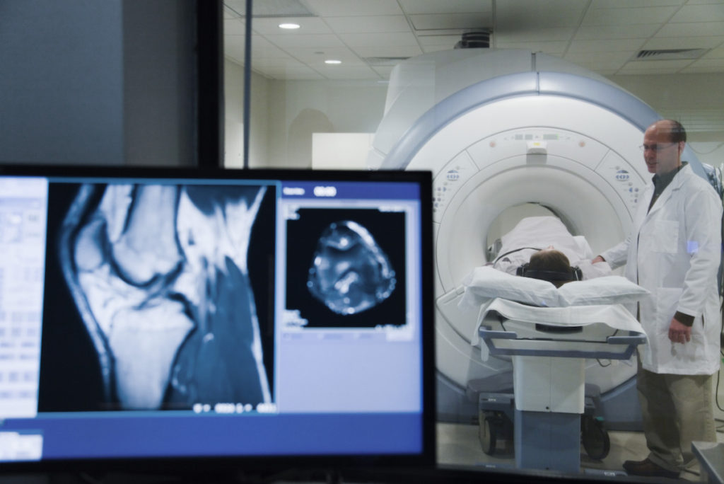
- Large Bore MRI
State-of-the-art magnetic resonance imaging (MRI) technology is available in the Imaging Center at Effingham Hosptial in Springfield. Our MRI provides high-quality images for physicians and is located in a convenient and pleasant setting for patients. If you would like an MRI, ask your doctor to refer you to Effingham Health System. Call 912-826-1400 for more information or to schedule an appointment.
- Positron Emission Tomography (PET Scan)
A positron emission tomography (PET) scan is an imaging test that allows your doctor to check for diseases in your body. The scan uses a special dye that has radioactive tracers. These tracers are injected into a vein in the arm, and organs and tissues then absorb the tracer.
- Radiography
Radiography is used to diagnose or treat patients by recording images of the internal structure of the body to assess the presence or absence of disease, foreign objects, and structural damage or anomaly.
During a radiographic procedure, an X-ray beam is passed through the body. A portion of the X-rays are absorbed or scattered by the internal structure and the remaining X-ray pattern is transmitted to a detector so that an image may be recorded for later evaluation. The recoding of the pattern may occur on film or through electronic means.
- Ultrasound
Ultrasound produces sound waves that are beamed into the body causing return echoes that are recorded to “visualize” structures beneath the skin. The ability to measure different echoes reflected from a variety of tissues allows a shadow picture to be constructed. The technology is especially accurate at seeing the interface between solid- and fluid-filled spaces. These are actually the same principles that allow sonar on boats to see the bottom of the ocean.